


“This is the first study to confirm the diagnostic utility of MRI for spinal pathology in SpA. Classic MRI SpA romanus lesions are also commonly seen in degenerative and malignant spines, but severe lesions in younger patients are highly specific for SpA. Collectively, these findings have broad implications for the diagnosis of SpA and for further studies into understanding patterns of disease expression.
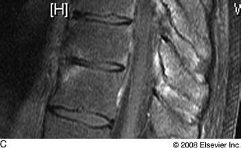
MRI shows typical Romanus ("shiny corner") lesions, representing anterior spondylitis
Dr. Bennett, who is at the Leeds Institute of Molecular Medicine, Leeds University, in the UK, led a study intended to determine if the reported classical lesions seen on MRI are specific to SpA or may represent other pathology. The researchers evaluated T1 and Fat suppressed MRI of the whole spine and SIJs on patients with back pain and on controls. They scored spine and SIJ bone marrow oedema (BMO) with the Leeds Scoring System, modified to assess severity of spinal and SIJ lesions. MRI-based diagnosis was compared to the gold standard treating physician diagnosis. The investigators analyzed the relationship between MRI romanus, posterior element and diffuse vertebral BMO lesions.
The study included 165 patients with back pain and 20 normal controls, mean age 53. The gold standard clinician diagnoses of these cases were SpA 35% (64), degenerative arthritis, 24% (45), malignancy 24% (45), normal 11% (20) or other 6% (11).
All subjects had whole spine and nearly half also had SIJ MRI. There was 74% agreement between study and gold standard diagnosis, and discrepancy was most common between SpA and degenerative arthritis.
Romanus lesions were common in all 3 major diagnoses. “Presence of any romanus lesion was of low diagnostic utility in SpA (sensitivity, 0.67 [95% CI 0.53 to 0.79]: specificity, 0.56 [0.47 to 0.65]),” Dr. Bennett said. “However, specificity increased to 0.82 (0.73 to 0.89) if >2 romanus lesions were present, to 0.87 (0.81 to 0.91) if severe romanus lesions were present, and to 1.00 (0.96 to 1.00) if severe romanus lesions were present in younger (ie, <51 years) patients. The presence/absence of degenerative discs did not change diagnostic utility.”
Any degree of posterior element bone marrow edema was highly specific for SpA (specificity, 0.87). This association was strongest if only mild or moderate lesions were present, and decreased if only severe lesions were present. This was due to the presence of high numbers of severe posterior element lesions in malignancy. Diffuse vertebral bone marrow edema was seen in 11% of SpA cases, 22% of degenerative cases, and 55% of malignant spines.
Dr. Bennett noted that 18% of SpA cases had normal SIJ scans, and 16% had normal spinal scans.
Active inflammation on MRI in the spine and sacroiliac joints in nonradiographic axial SpA
| Nonradiographic axial SpA, % (N = 160) |
AS, % (N=208) | P-value | |
| Patients with active sacroiliitis in MRI | 77.8 (112/144) | 76.7 (132/172) | P = .978 |
| Patients with active inflammation in the spine in MRI | 28.8 (17/59) | 56.3 (54/96) | P = .004 |
| Patients with isolated active inflammation in the spine in MRI and without sacroiliitis in MRI | 6.8 (3/44) | 10 (6/60) | P = .569 |
MRI = magnetic resonance imaging; SpA = spondyloarthritis; AS = ankylosing spondylitis.
Source: Adapted from Song I-H, et al.2
Sometimes sacroiliac MRI is all you need
In related work, In-Ho Song, MD, of Charité Medical University, in Berlin, Germany, found that among patients with nonradiographic axial SpA, only 6.8% had active inflammation in the spine but not active sacroiliitis.
“For establishing a diagnosis in suspected nonradiographic axial SpA, performing an MRI of the SIJ, irrespective of the clinical location of back pain, seems to be an efficient approach in daily clinical practice,” Dr. Song said.
This study was based on retrospective data from 362 patients seen in an ambulatory back pain clinic. Of these, 160 were classified as nonradiographic axial SpA or axial spondyloarthritis, and 202 as ankylosing spondylit5is. The nonradiographic axial SpA patients were significantly more likely to be female, not HLA-B27 positive, and have shorter disease duration than the AS patients (5.7 versus 11.2 years).
Frequency of active inflammation in the SIJ on MRI was similar in the nonradiographic axial SpA patients (77.8%) and AS patients (76.7%). Active inflammation in the spine was significantly less common in the non-radiographic axial SpA patients (28.8% versus 56.3%).
Among the 44 nonradiographic axial SpA patients who had undergone MRI of both the SIJ and the spine, only 3 (6.8%) had isolated active inflammation in the spine but not active sacroiliitis.
“There were 31 patients with nonradiographic axial SpA reporting spinal pain. Of these 31 patients, only 29.0% (9/31) had active inflammation in the spine on MRI while only 19.4% (6/31) had no active inflammation in the SIJ on MRI. “Active spinal inflammation without active sacroiliitis on MRI was found in only about 7% of patients with nonradiographic axial SpA,” Dr. Song concluded (see Table).
References
1. Bennett AN, Rheman A, Hensor EMA, et al. The diagnostic utility of MRI in spondyloarthropathy. American College of Rheumatology 2008 meeting, San Francisco, October 26, 2008. Presentation 510.
2. Song I-H, Hilgert E, Brandt HC, et al. Inflammatory lesions on magnetic resonance imaging in the spine and sacroiliac joints in patients with non-radiographic axial spondyloarthritis. Presented at: American College of Rheumatology 2008 meeting; San Francisco, Calif; October 26, 2008. Presentation 519.