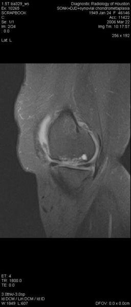
Subchondral cysts follow bone bruises
“Subchondral bone marrow edema-like lesions are strongly associated with subchondral cysts in the same subregion, supporting the bony contusion theory of SC formation.”—Michel D. Crema, MD
In the first study, Michel Crema, MD, et al reported that subchondral cysts in patients with knee OA are strongly associated with subchondral bone marrow edema-like lesions (BMLs), which indicates that these cysts likely follow bone contusions, not synovial fluid intrusion.
“Subchondral BMLs are strongly associated with SCs in the same subregion, supporting the bony contusion theory of SC formation,” Dr. Crema reported. “A substantial amount of SCs were located in subregions without full-thickness cartilage loss, which speaks against the synovial fluid intrusion theory. Our data supports the bony contusion theory of subchondral cyst formation in patients with or at risk for knee osteoarthritis.”
This study is part of the ongoing Multicenter Osteoarthritis Study (MOST). In this analysis the researchers used MRI to detect SCs, which present as well-defined rounded areas of fluid-like signal intensity on non-enhanced imaging. They then analyzed the association of SCs with subchondral bone marrow edema-like lesions (BMLs) and cartilage status in the same subregions where the SCs appeared. This allowed them to determine the roles played by bony contusion and by synovial fluid intrusion in causing subchondral cysts.
The MRI protocol included axial and sagittal proton-density weighted fat-suppressed fast spin echo sequences and a coronal STIR sequence. The investigators subdivided images of the tibiofemoral and patellofemoral joints into 14 subregions and scored SCs and BMLs from 0 to 3.
 Subchondral cysts precede cartilage loss
Subregions with SCs were graded for cartilage status as 0=normal, 1=partial-thickness loss, and 2=full-thickness loss. Dr. Crema assessed the cross-sectional association of BMLs and SCs in the same subregion using logistic regression, and determined SC distribution in subregions with normal cartilage, partial-thickness loss, and full-thickness loss of cartilage.
The investigators analyzed imaging data for 400 knees (5600 subregions). They found subchondral cysts in 260 subregions (4.6%) and BMLs in 757 subregions (13.5%).
Subchondral cysts were 83 times more common if the subregion had a BML [OR of 83.5 (95% CI 42.5-164)], and subchondral cysts became more common as BML size increased.
Furthermore, MRI identified subchondral cysts in 121 subregions (46.5%) that did not have full-thickness cartilage loss.
Bone marrow edema in tibia marks OA severity
In a related study, Jenanan Vairavamurthy, MD, and colleagues used a semi-quantitative marrow edema-based 3T MRI method to measure knee OA severity in 37 patients.
They compared MRI-detected changes in bone marrow edema to radiographically determined OA severity, evaluated as overall Kellgren and Lawrence (KL) grade on standard semi-flexed radiographs.
MRI included sagittal T2-weighted fat saturated spin-echo images (TR/TE=4000ms/75ms, FOV=15cm, matrix=256X128, slice thickness=3.0mm, receiver bandwidth 130Hz/pixel). Bone marrow edema (BME) volumes were quantified by location on T2-weighted fat saturated images, blinded to radiographic appearance.
Dr. Vairavamurthy reported that the averaged BME volume data for both medial femur and lateral femur showed a considerable scatter against the KL score, but the medial tibia and lateral tibia BME values closely tracked KL scores.
“Statistical analysis revealed no significant correlation between bone marrow edema and KL scores in the medial femoral, lateral femoral, and lateral tibial compartments...However, medial tibia showed a clear trend of increased amount of bone marrow edema with increasing KL score (r=0.35, P<0.0360). Changes in compartment-wise bone marrow edema (BME) measured by 3T MRI provide an important tool for assessing the severity of knee osteoarthritis in addition to radiography,” Dr. Vairavamurthy concluded.
References
1. Crema MD, Roemer F, Marra MD, et al. MRI-detected bone marrow edema-like lesions are strongly associated with subchondral cysts in patients with or at risk for knee osteoarthritis: The MOST Study. Presented at: Radiological Society of North America 2008 meeting; November 30, 2008-December 5, 2008; Chicago, Ill. Presentation No. SSA14-07.
2. Vairavamurthy J, Regatte R, Schweitzer M. A semi-quantitative marrow edema-based approach for assessing the severity of knee osteoarthritis (OA) using 3T MRI. Presented at: Radiological Society of North America 2008 meeting; November 30, 2008-December 5, 2008; Chicago, Ill. Presentation No. SSA14-05.


