


“Our research shows that it’s actually the strongest muscle in the back because of its unique design. It’s like a long, skinny pencil packed with millions of tiny fibers.”—Richard L. Lieber, PhD
The multifidus has been largely disregarded (and often disrupted) during invasive lumbar surgeries but is essential for keeping us upright and reducing pressure on our intervertebral discs, according to researchers at the University of California, San Diego School of Medicine. Their groundbreaking findings about the potentially important “scaffolding” role of the multifidus [FIGURE] were published online in advance of the January issue of the Journal of Bone and Joint Surgery.1Multifidus: strongest muscle in the back
 “The multifidus muscle was formerly thought to be relatively unimportant based on its fairly small size,” said senior author Richard L. Lieber, PhD, Professor and Vice Chair of UC San Diego’s Department of Orthopedic Surgery and Director of the National Center for Skeletal Muscle Rehabilitation Research, based at UC San Diego.
“Our research shows that it’s actually the strongest muscle in the back because of its unique design. It’s like a long, skinny pencil packed with millions of tiny fibers,” Dr. Lieber said.
The researchers discovered using cadaveric samples that the multifidus has a unique structure: short fibers arranged within rods. These fibers are stiffer than any other in the body. The researchers also used laser diffraction to measure muscle internal properties during back surgery. This showed that the multifidus’ unique design serves a critical function as a stabilizer of the lumbar spine.
Multifidus has critical implications for back surgery
These findings could have implications for surgery, according to Steven R. Garfin, MD, Professor and Chair of UCSD’s Department of Orthopaedic Surgery.
“It is important to identify what each individual muscle does, and this is just a start, showing that the multifidus contributes significantly to spinal stabilization,” Dr. Garfin said.
The lumbar vertebrae carry the most body weight and are subject to the most force and stress along the spine.
“The multifidus back muscle keeps us vertical and takes pressure off the discs,” said Dr. Lieber. “When muscle function is poor due to back problems, support is lost.”
Dr. Garfin explained that surgery to treat spinal disorders sometimes actually disrupts the multifidus, decreasing spinal stability and increasing lower back pain.
Multifidus: stronger when stretched
Dr. Lieber said that many muscles get weaker as they are extended, but the multifidus actually becomes stronger as it lengthens during spine flexion
“The length of the sarcomere—the structure within the muscle cell where filaments overlap to produce the movements required for muscle contraction—is shorter in the multifidus than in any other muscle cell,” explained lead author Samuel R. Ward, P.T., Ph.D., Assistant Professor of Radiology at UC San Diego School of Medicine. “But as it gets longer, for instance as a person leans forward, the multifidus actually strengthens.”
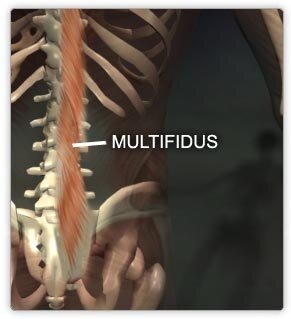
The multifidus consists of a number of fleshy and tendinous fasciculi, which fill the grooves on either side of the vertebral spinous processes from the sacrum to the axis. The very thin multifidus is deep in the spin, spans three joint segments, and works to stabilize the joints at each segmental level. The multifidus lies deep to the spinal erectors, transverse abdominus, and internal and external obliques.
The researchers used lumbar spines of 8 cadaver specimens, excised en bloc from T12 to the sacrum. They isolated multifidus muscles from each vertebral level and measured the mass, sarcomere length, normalized fiber length, physiological cross-sectional area, and fiber length-to-muscle ratio. They used intraoperative biopsy specimens from living patients to measure sarcomere lengths with the spine in flexion and extension.
"The architectural design (a high cross-sectional area and a low fiber length-to-muscle length ratio) demonstrates that the multifidus muscle is uniquely designed as a stabilizer to produce large forces. Furthermore, multifidus sarcomeres are positioned on the ascending portion of the length-tension curve, allowing the muscle to become stronger as the spine assumes a forward-leaning posture," the authors write.
Reference
1. Ward SR, Kim CW, Eng CM, et al. Architectural analysis and intraoperative measurements demonstrate the unique design of the multifidus muscle for lumbar spine stability. J Bone Joint Surg Am 2009;91:176-185.